Neural circuits that control REM sleep in mice identified
October 4, 2013
[+]
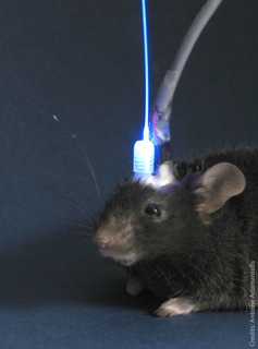
Scientists have controlled the duration of REM (rapid eye movements,
associated with dreaming) sleep in mice by activating
melanin-concentrating hormone (MCH)-expressing neurons in the lateral
hypothalamus, using optogenetics (controlling neuronal activity with
light).
Out like a light: laser light controls REM sleep in mice (credit: Douglas Institute)
“These research findings could help us better grasp how the brain controls sleep and better understand the role of sleep in humans. These results could also lead to new therapeutic strategies to treat sleep disorders along with associated neuropsychiatric problems,” said Dr. Antoine Adamantidis, who led the research.
He is an assistant professor at McGill University, a researcher at Douglas Institute, and is the Canada Research Chair in Neural Circuits and Optogenetics.
“This research will eventually lead to the identification of new therapeutic targets for treatment of sleep disorders (insomnia, fragmented sleep, etc.) as well as sleep disturbances associated with psychiatric disorders (such as major depression and schizophrenia),” Adamantidis explained to KurzweilAI.
[+]
“In a previous studies , we identified hypocretin/orexin (Adamantidis et al., Nature 2007) and norepinephrine (Carter et al., 2009 Nat. Neurosci.) neurons as important arousal circuits.”
Using
optogenetic laser light for acute activation of lateral hypothalamus
MCH neurons at the onset of REM sleep extended the duration of REM (but
not non-REM) sleep episodes (credit: Sonia Jego et al., Nature Neuroscience)
Researchers at Howard Hughes Medical Institute, Rockefeller University Laboratory of Molecular Genetics, MRC National Institute for Medical Research, and King’s College London where also involved.
Research funding was provided by Human Frontier Science Program, Canada Foundation for Innovation, Canadian Research Chair Program (Tier 2 Chair), Canadian Institutes of Health Research (CIHR), Natural Sciences and Engineering Research Council of Canada (NSERC), Fonds de Recherche du Québec – Santé (FRQS), McGill University, and Douglas Institute Foundation.
Abstract of Nature Neuroscience reference
Rapid-eye movement (REM) sleep correlates with neuronal activity in the brainstem, basal forebrain and lateral hypothalamus. Lateral hypothalamus melanin-concentrating hormone (MCH)-expressing neurons are active during sleep, but their effects on REM sleep remain unclear. Using optogenetic tools in newly generated Tg(Pmch-cre) mice, we found that acute activation of MCH neurons (ChETA, SSFO) at the onset of REM sleep extended the duration of REM, but not non-REM, sleep episodes. In contrast, their acute silencing (eNpHR3.0, archaerhodopsin) reduced the frequency and amplitude of hippocampal theta rhythm without affecting REM sleep duration. In vitro activation of MCH neuron terminals induced GABAA-mediated inhibitory postsynaptic currents in wake-promoting histaminergic neurons of the tuberomammillary nucleus (TMN), and in vivo activation of MCH neuron terminals in TMN or medial septum also prolonged REM sleep episodes. Collectively, these results suggest that activation of MCH neurons maintains REM sleep, possibly through inhibition of arousal circuits in the mammalian brain.
(¯`*• Global Source and/or more resources at http://goo.gl/zvSV7 │ www.Future-Observatory.blogspot.com and on LinkeIn Group's "Becoming Aware of the Futures" at http://goo.gl/8qKBbK │ @SciCzar │ Point of Contact: www.linkedin.com/in/AndresAgostini
 Washington