Evidence that dendrites actively process information in the brain
The brain's theoretical information processing power has just been multiplied
October 29, 2013
[+]
University of North Carolina at Chapel Hill researchers have discovered
that dendrites do more than passively relay information from one neuron
to the next — they actively process information, according to Spencer
Smith, PhD, an assistant professor in the UNC School of Medicine.
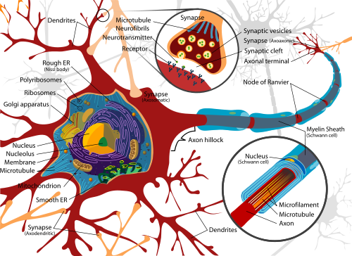
Diagram of a typical neuron. Red branches: dentrites; blue segments: axon. (Credit: Wikimedia Commons)
Axons are where neurons conventionally generate electrical spikes, but many of the same molecules that support axonal electrical spikes (firing) are also present in dendrites.
Previous research using dissected brain tissue had demonstrated that dendrites can use those molecules to generate electrical spikes themselves, but it was unclear whether normal brain activity uses those dendritic spikes.
For example, could dendritic spikes be involved in how we see?
The evidence
[+]
To find out, the researchers used a two-photon microscope system and
they used patch-clamp electrophysiology to attach a microscopic glass
pipette electrode, filled with a physiological solution, to a neuronal
dendrite in the brain of a mouse.
A
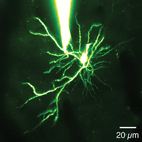
pipette (white object at top) attached to a dendrite in the brain of a
mouse, allowing researchers to measure electrical activity at a
dendritic spike in the mouse primary visual
cortex (credit: University of North Carolina at Chapel Hill)
cortex (credit: University of North Carolina at Chapel Hill)
As the mice viewed visual stimuli on a computer screen, the researchers saw an unusual pattern of electrical signals — bursts of spikes — in the dendrite.
Smith’s team then found that the dendritic spikes occurred selectively, depending on the visual stimulus, indicating that the dendrites in fact processed information about what the animal was seeing.
To provide visual evidence of their finding, Smith’s team filled neurons with calcium dye, which provided an optical readout of spiking.
This revealed that dendrites fired spikes while other parts of the neuron did not, meaning that the spikes were the result of local processing within the dendrites — not a electrical signal from the body of the neuron.
[+]
Study co-author Tiago Branco, PhD, created a biophysical,
mathematical model of neurons and found that known mechanisms could
support the dendritic spiking recorded electrically, further validating
the interpretation of the data.
Schematic of the recording and imaging setup for in vivo
dendritic recordings of responses to visual stimulation (left) using a
patch clamp (rod) and two-photon microscope (top) (credit: Spencer L.
Smith et al./Nature)
The team plans to explore what this newly discovered dendritic role may play in brain circuitry, particularly in conditions like Timothy syndrome, in which the integration of dendritic signals may go awry.
This work was supported by a Long-Term Fellowship and a Career Development Award from the Human Frontier Science Program, and a Klingenstein Fellowship to S. Smith, a Helen Lyng White Fellowship to I. Smith, a Wellcome Trust and Royal Society Fellowship, and Medical Research Council (UK) support to T. Branco, and grants from the Wellcome Trust, the European Research Council, and Gatsby Charitable Foundation to M. Häusser.
Abstract of Nature paper
Neuronal dendrites are electrically excitable: they can generate regenerative events such as dendritic spikes in response to sufficiently strong synaptic input. Although such events have been observed in many neuronal types, it is not well understood how active dendrites contribute to the tuning of neuronal output in vivo. Here we show that dendritic spikes increase the selectivity of neuronal responses to the orientation of a visual stimulus (orientation tuning). We performed direct patch-clamp recordings from the dendrites of pyramidal neurons in the primary visual cortex of lightly anaesthetized and awake mice, during sensory processing. Visual stimulation triggered regenerative local dendritic spikes that were distinct from back-propagating action potentials. These events were orientation tuned and were suppressed by either hyperpolarization of membrane potential or intracellular blockade of NMDA (N-methyl-D-aspartate) receptors. Both of these manipulations also decreased the selectivity of subthreshold orientation tuning measured at the soma, thus linking dendritic regenerative events to somatic orientation tuning. Together, our results suggest that dendritic spikes that are triggered by visual input contribute to a fundamental cortical computation: enhancing orientation selectivity in the visual cortex. Thus, dendritic excitability is an essential component of behaviourally relevant computations in neurons.
(¯`*• Global Source and/or more resources at http://goo.gl/zvSV7 │ www.Future-Observatory.blogspot.com and on LinkeIn Group's "Becoming Aware of the Futures" at http://goo.gl/8qKBbK │ @SciCzar │ Point of Contact: www.linkedin.com/in/AndresAgostini
 Washington