Detecting objects as small as protein molecules using multispectral imaging
December 10, 2013
[+]
Richard Martel and his research team at the Department of Chemistry of the University of Montreal
have discovered a method to improve detection of the “infinitely small”
by encapsulating a dye inside carbon nanotubes for multispectral
imaging.
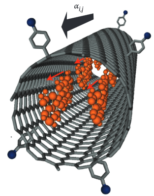
A nanoprobe, consisting of a dye encapsulated in a carbon nanotube (credit: Universite de Montreal)
“Raman scattering provides information on the ways molecules vibrate, which is equivalent to taking their fingerprint. It’s a bit like a bar code,” said Martel. “Raman signals are specific for each molecule and thus useful in identifying these molecules.”
The discovery by Martel’s team is that Raman scattering of dye-nanotube nanoprobes is so effective that a single particle of this type can be located and identified. All one needs is an optical scanner capable of detecting this nanoprobe, much like a fingerprint.
“By incorporating these nanoparticles in an object, you can make it perfectly traceable,” he said. Due to their unique structure, carbon nanotubes, which are electrically conductive, can be used as containers for various molecules.
Coupled with a dye, these nanoprobes can increase the complexity and strength of the received signal. They are about one nanometer (nm) in diameter and 500 nm long, yet they send a Raman signal one million times stronger than the other molecules in the surrounding material.
Applications: from diseased cells to preventing counterfeit money
According to Martel, the applications from this “giant Raman scattering” discovery are numerous. In medicine, nanoprobes could lead to improved diagnostics and better treatment by adhering to the surface of diseased cells. These specifically modified nanoprobes could, in effect, be grafted to bacteria or even proteins, allowing them to be easily identified.
One could also imagine custom officers scanning passports with Raman multispectral mode (involving several signals). Nanoprobes could also be used in banknote ink, making counterfeiting virtually impossible.
The beauty of it, said Martel, is that the phenomenon is generalized, and many types of dyes can be used to make nanoprobes or tags, whose “bar codes” are all different. “So far, more than 10 different tags have been made. We could, in theory, create as many of these tags as there are bacteria and use this principle to identify them with a microscope operating in Raman mode.”
The story of Raman signals
Raman scattering mode is an optical phenomenon discovered in 1928 by the physicist Chandrasekhara Venkata Raman. The effect involves the inelastic scattering of photons, i.e. the physical phenomenon by which a medium can modify the frequency of the light impinging on it.
The difference corresponds to an exchange of energy (wavelength) between the light beam and the medium. In this way, scattered light does not have the same wavelength as incidental light. The technique has become widely used since the advent of the laser in the industry and for research .
But until now, molecular Raman signals have been too weak to serve the needs of optical imaging effectively. So researchers have used other more sensitive techniques but which are less specific because they have no “bar code.” “It is technically possible, however, to enhance the Raman signals of molecules using rough metallic surfaces,” said Martel. “But their sizes limit the applications of Raman spectroscopy and imaging.”
By aligning dye molecules encapsulated in carbon nanotubes, the researchers were able to amplify the Raman signals of these molecules, which until now have not been strong enough to detect.
Their discovery is presented in the November 24 online edition of the journal Nature Photonics.
Besides Richard Martel, E. Gaufrès, N. Y. Wa Tang, F. Lapointe, J. Cabana, M. A. Nadon, N. Cottenye, F. Raymond, all of the Université de Montréal, and T. Szkopek, University McGill, contributed to this discovery.
Abstract of Nature Photonics paper
Raman spectroscopy uses visible light to acquire vibrational fingerprints of molecules, thus making it a powerful tool for chemical analysis in a wide range of media. However, its potential for optical imaging at high resolution is severely limited by the fact that the Raman effect is weak. Here, we report the discovery of a giant Raman scattering effect from encapsulated and aggregated dye molecules inside single-walled carbon nanotubes. Measurements performed on rod-like dyes such as α-sexithiophene and β-carotene, assembled inside single-walled carbon nanotubes as highly polarizable J-aggregates, indicate a resonant Raman cross-section of (3 ± 2) × 10−21 cm2 sr−1, which is well above the cross-section required for detecting individual aggregates at the highest optical resolution. Free from fluorescence background and photobleaching, this giant Raman effect allows the realization of a library of functionalized nanoprobe labels for Raman imaging with robust detection using multispectral analysis.
(¯`*• Global Source and/or more resources at http://goo.gl/zvSV7 │ www.Future-Observatory.blogspot.com and on LinkeIn Group's "Becoming Aware of the Futures" at http://goo.gl/8qKBbK │ @SciCzar │ Point of Contact: www.linkedin.com/in/AndresAgostini
 Washington