QUANTA

Friday, October 4, 2013

Neural circuits that control REM sleep in mice identified

October 4, 2013
[+]
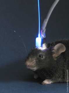
Out like a light: laser light controls REM sleep in mice (credit: Douglas Institute)
Scientists have controlled the duration of REM (rapid eye movements, associated with dreaming) sleep in mice by activating melanin-concentrating hormone (MCH)-expressing neurons in the lateral hypothalamus, using optogenetics (controlling neuronal activity with light).
“These research findings could help us better grasp how the brain controls sleep and better understand the role of sleep in humans. These results could also lead to new therapeutic strategies to treat sleep disorders along with associated neuropsychiatric problems,” said Dr. Antoine Adamantidis, who led the research.
He is an assistant professor at McGill University, a researcher at Douglas Institute, and is the Canada Research Chair in Neural Circuits and Optogenetics.
“This research will eventually lead to the identification of new therapeutic targets for treatment of sleep disorders (insomnia, fragmented sleep, etc.) as well as sleep disturbances associated with psychiatric disorders (such as major depression and schizophrenia),” Adamantidis explained to KurzweilAI.
[+]
Using optogenetic laser light for acute activation of lateral hypothalamus MCH neurons at the onset of REM sleep extended the duration of REM (but not non-REM) sleep episodes (credit: Sonia Jego et al., Nature Neuroscience)
“In a previous studies , we identified hypocretin/orexin (Adamantidis et al., Nature 2007) and norepinephrine (Carter et al., 2009 Nat. Neurosci.) neurons as important arousal circuits.”
Researchers at Howard Hughes Medical Institute, Rockefeller University Laboratory of Molecular Genetics, MRC National Institute for Medical Research, and King’s College London where also involved.
Research funding was provided by Human Frontier Science Program, Canada Foundation for Innovation, Canadian Research Chair Program (Tier 2 Chair), Canadian Institutes of Health Research (CIHR), Natural Sciences and Engineering Research Council of Canada (NSERC), Fonds de Recherche du Québec – Santé (FRQS), McGill University, and Douglas Institute Foundation.

Abstract of Nature Neuroscience reference

Rapid-eye movement (REM) sleep correlates with neuronal activity in the brainstem, basal forebrain and lateral hypothalamus. Lateral hypothalamus melanin-concentrating hormone (MCH)-expressing neurons are active during sleep, but their effects on REM sleep remain unclear. Using optogenetic tools in newly generated Tg(Pmch-cre) mice, we found that acute activation of MCH neurons (ChETA, SSFO) at the onset of REM sleep extended the duration of REM, but not non-REM, sleep episodes. In contrast, their acute silencing (eNpHR3.0, archaerhodopsin) reduced the frequency and amplitude of hippocampal theta rhythm without affecting REM sleep duration. In vitro activation of MCH neuron terminals induced GABAA-mediated inhibitory postsynaptic currents in wake-promoting histaminergic neurons of the tuberomammillary nucleus (TMN), and in vivo activation of MCH neuron terminals in TMN or medial septum also prolonged REM sleep episodes. Collectively, these results suggest that activation of MCH neurons maintains REM sleep, possibly through inhibition of arousal circuits in the mammalian brain.


(¯`*• Global Source and/or more resources at http://goo.gl/zvSV7 │ www.Future-Observatory.blogspot.com and on LinkeIn Group's "Becoming Aware of the Futures" at http://goo.gl/8qKBbK │ @SciCzar │ Point of Contact: www.linkedin.com/in/AndresAgostini

Cells from small biopsies can be used to grow large numbers of a patient’s own protective brain cells

October 4, 2013
[+]
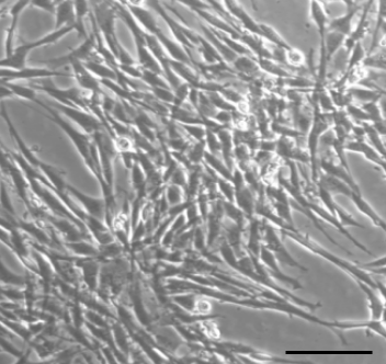
Cells from brain biopsy for patient with Parkinson’s disease.Scale bar: 50 microns. (Credit: Hu Xu et al./The FASEB Journal)
Scientists at the University of Western Ontario in Canada enrolled patients with Parkinson’s disease who were scheduled to have deep brain stimulation (DBS) surgery and removed small biopsies near the surface of the brain.
They then multiplied the cells in culture to generate millions of patient-specific cells that were then subjected to genetic analysis.
These cells exhibited regeneration and characteristics of a fundamental class of brain cells, called glia. The cells expressed a broad array of natural and potent protective agents, called neurotrophic factors.
“It is our hope that the results of this study provide a footing for further advancement of personalized, cell-based treatments for currently incurable and devastating neurological disorders,” said Matthew O. Hebb, M.D., Ph.D., FRCSC, a researcher involved in the work from the Departments of Clinical Neurological Sciences (Neurosurgery), Oncology and Otolaryngology at the University of Western Ontario in Canada.
Such lab-grown therapeutic brain cells could be used in the future to treat a wide range of neurological conditions like Parkinson’s, and also express a broad array of natural and potent protective agents providing preservation and protection against injury, toxins and diseases.


(¯`*• Global Source and/or more resources at http://goo.gl/zvSV7 │ www.Future-Observatory.blogspot.com and on LinkeIn Group's "Becoming Aware of the Futures" at http://goo.gl/8qKBbK │ @SciCzar │ Point of Contact: www.linkedin.com/in/AndresAgostini
MIT inventor unleashes hundreds of self-assembling cube swarmbots

October 4, 2013
[+]
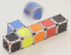
Self-assembling swarming microbots (credit: MIT)
The experts said it couldn’t be done. But research scientist John Romanishin of MIT’s Computer Science and Artificial Intelligence Laboratory (CSAIL) has created  M-Blocks — cube robots with no external moving parts.
Despite that, they can magically climb over and around one another, leap through the air, roll across the ground, snap together into different shapes, and even move while suspended upside down from metallic surfaces.
Self-assembling swarmbots
Imagine hordes of swarming microbots that can self-assemble, like the “liquid steel” androids in the movie “Terminator II.”
Armies of these mobile cubes could temporarily repair bridges or buildings during emergencies. These cubes could assemble into different types of furniture or heavy equipment as needed. And they could swarm into environments hostile or inaccessible to humans, diagnose problems, and then reorganize themselves to provide solutions.
They could even be special-purpose cubes: containing cameras, or lights, or battery packs, or other equipment that the mobile cubes could transport.
How M-Blocks work
mit_modular_robot
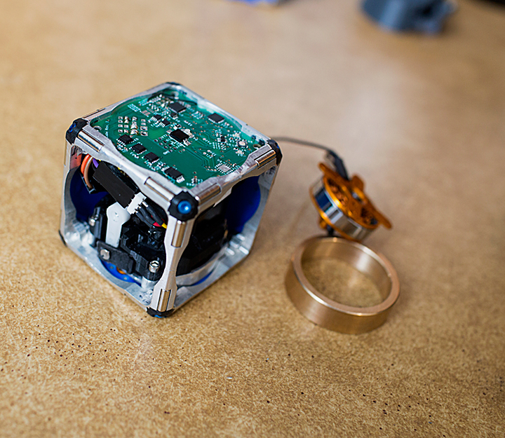
A prototype of a new modular robot, with its innards exposed and its flywheel — which gives it the ability to move independently — pulled out (credit: M. Scott Brauer, MIT)
The trick: a flywheel that can reach speeds of 20,000 revolutions per minute. When the flywheel is braked, it imparts its angular momentum to the cube. And on each edge of an M-Block and on every face, cleverly arranged permanent magnets allow any two cubes to attach to each other.
To compensate for its static instability, the researchers’ robot relies on some ingenious engineering. On each edge of a cube are two cylindrical magnets, mounted like rolling pins.
When two cubes approach each other, the magnets naturally rotate, so that north poles align with south, and vice versa. Any face of any cube can thus attach to any face of any other.
The cubes’ edges are also beveled, so when two cubes are face to face, there’s a slight gap between their magnets. When one cube begins to flip on top of another, the bevels, and thus the magnets, touch. The connection between the cubes becomes much stronger, anchoring the pivot. On each face of a cube are four more pairs of smaller magnets, arranged symmetrically, which help snap a moving cube into place when it lands on top of another.
A cube army
The MIT researchers are currently building an army of 100 cubes, each of which can move in any direction, and designing algorithms to guide them. “We want hundreds of cubes, scattered randomly across the floor, to be able to identify each other, coalesce, and autonomously transform into a chair, or a ladder, or a desk, on demand,” Romanishin says.
Romanishin, robotics professor Daniela Rus, and postdoc Kyle Gilpin will present a paper describing their new robots at the IEEE/RSJ International Conference on Intelligent Robots and Systems in Japan in November.


(¯`*• Global Source and/or more resources at http://goo.gl/zvSV7 │ www.Future-Observatory.blogspot.com and on LinkeIn Group's "Becoming Aware of the Futures" at http://goo.gl/8qKBbK │ @SciCzar │ Point of Contact: www.linkedin.com/in/AndresAgostini
Thursday, October 3, 2013

’4D printing’ adaptive materials

October 3, 2013
[+]
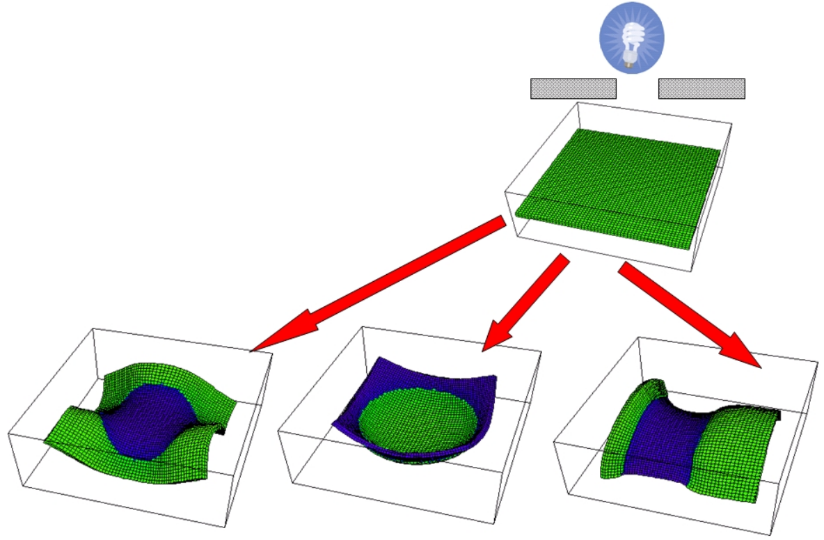
Example of a programmable 4D material: photo-morphing of initially flat hydrogel sample into various shapes by illumination (credit: Anna C. Balazs)
Researchers from three universities are proposing to add a dimension to 3D printing by developing “4D” materials that can exhibit behavior that changes over time.
Imagine an automobile coating that changes its structure to adapt to a humid environment or a salt-covered road, better protecting the car from corrosion. Or consider a soldier’s uniform that could alter its camouflage or more effectively protect against poison gas or shrapnel upon contact.
With an $855,000 three-year grant from the United States Army Research Office, the researchers from the University of Pittsburgh’s Swanson School of Engineering, Harvard School of Engineering and Applied Sciences, and the University of Illinois plan to manipulate materials at nano and micro levels.
The goal: produce, via 3D printing, materials that can modify their structures over time at the macro level.
Adaptive biomimetic composites
“Rather than construct a static material or one that simply changes its shape, we’re proposing the development of adaptive, biomimetic composites that reprogram their shape, properties, or functionality on demand, based upon external stimuli,” said principal investigator Anna C. Balazs, the Robert v. d. Luft Distinguished Professor of Chemical Engineering in Pitt’s Swanson School of Engineering and a researcher in the computational design of chemo-mechanically responsive gels and composites.
“By integrating our abilities to print precise, three-dimensional, hierarchically-structured materials; synthesize stimuli-responsive components; and predict the temporal behavior of the system, we expect to build the foundation for the new field of 4D printing.”
Co-investigators are Jennifer A. Lewis, the Hansjörg Wyss Professor of Biologically Inspired Engineering at the Harvard School of Engineering and Applied Sciences and an expert in 3D printing of functional materials; and Ralph G. Nuzzo, the G. L. Clark Professor of Chemistry and Professor of Materials Science and Engineering at the University of Illinois, a synthetic chemist who has created novel stimuli-responsive materials.
Lewis added that current 3D printing technology allows the researchers to build in complicated functionality at the nano and micro levels within specific areas of the structure. “Composites that can be reconfigured in the presence of different stimuli could dramatically extend the reach of 3D printing.”
The research will use responsive fillers embedded within a stimuli-responsive hydrogel. Nuzzo says this opens new routes for producing the next generation of smart sensors, coatings, textiles, and structural components.
“The ability to create one fabric that responds to light by changing its color, and to temperature by altering its permeability, and even to an external force by hardening its structure, becomes possible through the creation of responsive materials that are simultaneously adaptive, flexible, lightweight, and strong. It’s this ‘complicated functionality’ that makes true 4D printing a game changer.”

(¯`*• Global Source and/or more resources at http://goo.gl/zvSV7 │ www.Future-Observatory.blogspot.com and on LinkeIn Group's "Becoming Aware of the Futures" at http://goo.gl/8qKBbK │ @SciCzar │ Point of Contact: www.linkedin.com/in/AndresAgostini
The secret of longevity for the world’s longest-living rodent: better protein creation

October 3, 2013
[+]
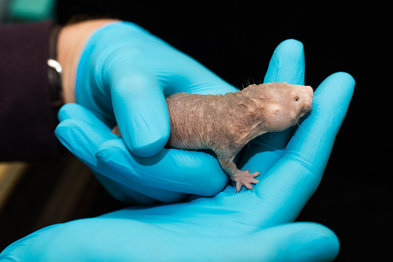
Naked mole rats are small, hairless, subterranean rodents native to eastern Africa (credit: Adam Fenster/University of Rochester)
Better-constructed proteins could explain why naked mole rats live long lives — about 30 years — and stay healthy until the very end, resisting cancer, say University of Rochester biologists Vera Gorbunova and Andrei Seluanov.
Their work focuses on naked mole rat ribosomes, which assemble amino acids into proteins. Ribosomes are composed of ribosomal RNA (rRNA) molecules and proteins.
When the ribosome connects amino acids together to create a protein, a mistake is occasionally introduced when an incorrect amino acid is inserted. But the researchers found that the proteins made by naked mole rat cells are up to 40 times less likely to contain such mistakes than the proteins made by mouse cells.
Gorbunova and Seluanov discovered a possible reason: the naked mole rat’s rRNA is unique.
Unique rRNA pattern
[+]
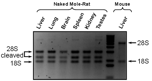
Naked mole rat rRNA in this NMR image shows an unusual pattern with cleaved 28S pattern: 3 bands, instead of 2 with mouse (credit: Jorge Azpurua et al./PNAS)
After applying a dye to a sample, they studied it under ultraviolet light. They found three dark bands — representing concentrations of different rRNA molecules — not the two bands that are characteristic of all other animals, suggesting that there is a “hidden break” in the naked mole rat rRNA.
When its rRNA strands cleave (split) at two specific locations, instead of floating off on their own, two remaining pieces from each strand stay close to each other. They act as a scaffold on which ribosomal proteins are assembled to create a functional ribosome, preventing aberrations. “This is important because proteins with no aberrations help the body to function more efficiently,” said Seluanov.
The next step for the biologists is to split mouse rRNA in the same way to see if it would lead to improved protein creation.
The two biologists hope their work will eventually result in pharmaceutical treatments that modulate protein synthesis in humans, though any medical solution is a long way off, they say.

Abstract for PNAS paper
The naked mole-rat (Heterocephalus glaber) is a subterranean eusocial rodent with a markedly long lifespan and resistance to tumorigenesis. Multiple data implicate modulation of protein translation in longevity. Here we report that 28S ribosomal RNA (rRNA) of the naked mole-rat is processed into two smaller fragments of unequal size. The two breakpoints are located in the 28S rRNA divergent region 6 and excise a fragment of 263 nt. The excised fragment is unique to the naked mole-rat rRNA and does not show homology to other genomic regions. Because this hidden break site could alter ribosome structure, we investigated whether translation rate and amino acid incorporation fidelity were altered. We report that naked mole-rat fibroblasts have significantly increased translational fidelity despite having comparable translation rates with mouse fibroblasts. Although we cannot directly test whether the unique 28S rRNA structure contributes to the increased fidelity of translation, we speculate that it may change the folding or dynamics of the large ribosomal subunit, altering the rate of GTP hydrolysis and/or interaction of the large subunit with tRNA during accommodation, thus affecting the fidelity of protein synthesis. In summary, our results show that naked mole-rat cells produce fewer aberrant proteins, supporting the hypothesis that the more stable proteome of the naked mole-rat contributes to its longevity.


(¯`*• Global Source and/or more resources at http://goo.gl/zvSV7 │ www.Future-Observatory.blogspot.com and on LinkeIn Group's "Becoming Aware of the Futures" at http://goo.gl/8qKBbK │ @SciCzar │ Point of Contact: www.linkedin.com/in/AndresAgostini
Seeing cells through silicon microfluidic devices

October 3, 2013
[+]
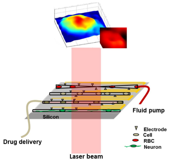
Schematic of the lab-on-chip system for the study of
mechanical, chemical, and electric perturbation of different types of cells
on silicon-based microfluidic and multi-electrode array platform.
Quantitative phase image (through silicon) of a human embryonic kidney cell and red blood cell  is shown on the top. (Credit: B. Joshi et al./Scientific Reports)
Scientists at MIT and the University of Texas at Arlington (UTA) have developed a new type of microscopy that can image cells through a silicon wafer, allowing them to precisely measure the size and mechanical behavior of cells behind the wafer.
The new technology, which relies on near-infrared light, could help scientists learn more about diseased or infected cells as they flow through silicon microfluidic devices.
“This has the potential to merge research in cellular visualization with all the exciting things you can do on a silicon wafer,” says Ishan Barman, a former postdoc in MIT’s Laser Biomedical Research Center (LBRC) and one of the lead authors of a paper describing the technology in the Oct. 2 issue of the journal Scientific Reports (open access).
The senior author is Samarendra Mohanty, an assistant professor of physics at UTA.
Silicon is commonly used to build “lab-on-a-chip” microfluidic devices, which can sort and analyze cells based on their molecular properties. Such devices have many potential applications in research and diagnostics, but they could be even more useful if scientists could image the cells inside the devices, says Barman, who is now an assistant professor of mechanical engineering at Johns Hopkins University.
To achieve that, Barman and colleagues took advantage of the fact that silicon is transparent to infrared and near-infrared wavelengths of light. They adapted a microscopy technique known as quantitative phase imaging, which works by sending a laser beam through a sample, then splitting the beam into two. By recombining those two beams and comparing the information carried by each one, the researchers can determine the sample’s height and its refractive index — a measure of how much the material forces light to bend as it passes through.
Traditional quantitative phase imaging uses a helium neon laser, which produces visible light, but for the new system the researchers used a titanium sapphire laser that can be tuned to infrared and near-infrared wavelengths (they found that light with a wavelength of 980 nanometers worked best).
Using this system, the researchers measured changes in the height of red blood cells, with nanoscale sensitivity, through a silicon wafer similar to those used in most electronics labs.
Examples of uses:
  • As red blood cells flow through the body, they often have to squeeze through very narrow vessels. When these cells are infected with malaria, they lose this ability to deform, and form clogs in tiny vessels. The new microscopy technique could help scientists study how this happens, the researchers say. It could also be used to study the dynamics of the malformed blood cells that cause sickle cell anemia.
  • The researchers also used their new system to monitor human embryonic kidney cells as pure water was added to their environment — a shock that forces the cells to absorb water and swell up. The researchers were able to measure how much the cells distended and calculate the change in their index of refraction.
  • Mohanty’s lab at UTA is now using the system to study how neurons grown on a silicon wafer communicate with each other.
“Nobody has shown this kind of microscopy of cellular structures before through a silicon substrate,” Mohanty says.
“This is an exciting new direction that is likely to open up enormous opportunities for quantitative phase imaging,” says Gabriel Popescu, an assistant professor of electrical engineering and computer science at the University of Illinois at Urbana-Champaign who was not part of the research team. “The possibilities are endless: From micro- and nanofluidic devices to structured substrates, the devices could target applications ranging from molecular sensing to whole-cell characterization and drug screening in cell populations,” he says.
In the Scientific Reports paper, the researchers used silicon wafers that were about 150 to 200 microns thick, but they have since shown that thicker silicon can be used if the wavelength of light is increased into the infrared range. The researchers are also working on modifying the system so that it can image in three dimensions, similar to a CT scan.
Other lead authors of the paper are former MIT postdoc Narahara Chari Dingari and UTA graduate students Bipin Joshi and Nelson Cardenas. Other authors are former MIT postdoc Jaqueline Soares, currently an assistant professor at Federal University of Ouro Preto, Brazil, and Ramachandra Rao Dasari, associate director of the LBRC.
The research was funded by the National Institute of Biomedical Imaging and Bioengineering and Nanoscope Technologies, LLC.
Department of Chemistry
“MIT shows how blood cells change shape”

Abstract for Scientific Reports paper
We report a novel technique for label-free, rapid visualization of structure and dynamics of live cells with nanoscale sensitivity through traditionally opaque media. Specifically, by combining principles of near-infrared (NIR) spectroscopy and quantitative phase imaging, functional characterization of cellular structure and dynamics through silicon substrates is realized in our study. We demonstrate the efficacy of the new approach by full-field imaging of erythrocyte morphology in their native states with a nm path length sensitivity. Additionally, we observe dynamic variations of human embryonic kidney cells, through a silicon substrate, in response to hypotonic stimulation with ms temporal resolution that also provides unique insight into the underlying biophysical changes. The proposed technology is fundamentally suited for high-performance investigations of biological specimens and significantly expands the options for visualization in complex microfluidic devices fabricated on silicon.


(¯`*• Global Source and/or more resources at http://goo.gl/zvSV7 │ www.Future-Observatory.blogspot.com and on LinkeIn Group's "Becoming Aware of the Futures" at http://goo.gl/8qKBbK │ @SciCzar │ Point of Contact: www.linkedin.com/in/AndresAgostini
Is ‘massive open online research’ (MOOR) the next frontier for education?

"The goal of this class is to make you fall in love with bioinformatics" --- Prof. Pavel Pevzner
October 3, 2013
UC San Diego is launching the first major online course that prominently features “massive open online research” (MOOR).
In “Bioinformatics Algorithms — Part 1,” UC San Diego computer science and engineering professor Pavel Pevzner and his graduate students are offering a course on Coursera that combines research with a MOOC (massive open online course) for the first time.
“All students who sign up for the course will be given an opportunity to work on specific research projects under the leadership of prominent bioinformatics scientists from different countries, who have agreed to interact and mentor their respective teams.”
“The natural progression of education is for people to make a transition from learning to research, which is a huge jump for many students, and essentially impossible for students in isolated areas,” said Ph.D. student Phillip Compeau, who helped develop the online course. “By integrating the research with an interactive text and a MOOC, it creates a pipeline to streamline this transition.”
Bioinformatics Algorithms (Part I) will run for eight weeks starting October 21, and students are now able to sign up and download some of the course materials. It is offered free of charge to everyone.
Book, content delivery system
Bioinf_coursera_part1
Bioinformatics Algorithms (Part 1) (credit: UCSD/Coursera)
Another unique feature of the online course: Pevzner and Compeau have developed Bioinformatics Algorithms: An Active-Learning Approach, a e-book supporting the course, while Pevzner’s colleagues in Russia developed a content delivery system that integrates the e-book with hundreds of quizzes and dozens of homework problems.
The U.S.-Russian team, led by Pevzner’s foreign student Nikolay Vyahhi, also implemented the online course using the beta version of Stepic, a new, fully integrated educational platform and startup developed by Vyahhi. (Stepic derives its name from the “step-by-step, epic” solution its developers delivered for electronic publishing.)
The course also provides access to Rosalind, a free online resource for learning bioinformatics through problem solving. Rosalind was developed by Pevzner’s students and colleagues in San Diego and St. Petersburg with funding from the Howard Hughes Medical Institute, the Russian Ministry of Education, and Russian Internet billionaires Yuri Milner and Pavel Durov through their “Start Fellows” award. Rosalind already has over 10,000 active users worldwide.
Rosalind — named in honor of British scientist Rosalind Franklin, whose X-ray crystallography with Raymond Gosling facilitated the discovery of the DNA double helix by Watson and Crick — will grade the programming assignments. They come in the form of bioinformatics problems of growing complexity as the course progresses.
“We developed Rosalind to inspire both biologists and computer science students,” said Rosalind principal developer Vyahhi, who worked with Pevzner during the latter’s sabbatical in Russia. “The platform allows biologists to develop vital programming skills for bioinformatics at their own pace, and Rosalind can also appeal to programmers who have never been exposed to some of the exciting computational problems generated by molecular biology.”
“We have already started testing the first modules of the Bioinformatics Algorithms course in top Russian universities,” said Pevzner. “We are getting good feedback, and we hope to reach out to early students registering for the Coursera course so we can perform a ‘stress’ test — just to make sure that we will be able to deliver on the promise of massive open online research projects, whether it’s hundreds or thousands of students who enroll.”
The UC San Diego course, produced in Calit2’s Qualcomm Institute at UC San Diego, will cover many algorithms underlying fundamental topics in bioinformatics and will try to answer questions like, “What DNA Patterns Play the Role of Molecular Clocks Inside Cells,” or, “How Have Humans Populated the Earth?” To answer these questions, Pevzner and his team will introduce fundamental computational concepts such as greedy and randomized algorithms, graph theory and combinatorics, dynamic programming and combinatorial pattern matching, and many others.
‘We want you to switch to bioinformatics’
“Whatever you are studying now — computer science, biology, or mathematics — we want you to switch to bioinformatics,” exhorted Pevzner in the promotional video. “Not because these disciplines are not important, quite the opposite; but because bioinformatics is so cool, and because it is built on all of these disciplines!”
Pevzner was working on his Ph.D. in discrete mathematics when he had his first encounter with bioinformatics, and he says that he started over in the “futuristic new discipline — and never looked back.” He is now a professor of Computer Science and Engineering at UC San Diego, where he holds the Ronald R. Taylor Chair.
In 2006, Pevzner was named a Howard Hughes Medical Institute Professor. His research has focused on the creation of bioinformatics algorithms for analyzing genome rearrangements, DNA sequencing, and computational proteomics. He is a Fellow of both the Association for Computing Machinery (ACM) and the International Society for Computational Biology (ISCB).
In 2011, Pevzner founded the Algorithmic Biology Laboratory in St. Petersburg, Russia, which develops the Rosalind bioinformatics platform.
Topping a long list of publications, Pevzner authored Computational Molecular Biology (The MIT Press, 2000), co-authored (jointly with Neil Jones) An Introduction to Bioinformatics Algorithms (The MIT Press, 2004), and co-edited (with Ron Shamir) Bioinformatics for Biologists (Cambridge University Press, 2011).
But asked about which book makes him most proud — and Pevzner is unequivocal. “Absolutely this new book for the Bioinformatics Algorithms course,” he said. “Thanks to Coursera and the online learning explosion, we have the opportunity to reach many more students than would be possible at one university or even one country. There are students everywhere who could use this as a stepping stone to a career in a field that is going to change the world as we know it. Who wouldn’t want to be a part of that?”


(¯`*• Global Source and/or more resources at http://goo.gl/zvSV7 │ www.Future-Observatory.blogspot.com and on LinkeIn Group's "Becoming Aware of the Futures" at http://goo.gl/8qKBbK │ @SciCzar │ Point of Contact: www.linkedin.com/in/AndresAgostini
Wednesday, October 2, 2013


New programming language directs DNA to build custom-designed molecules

October 2, 2013
[+]
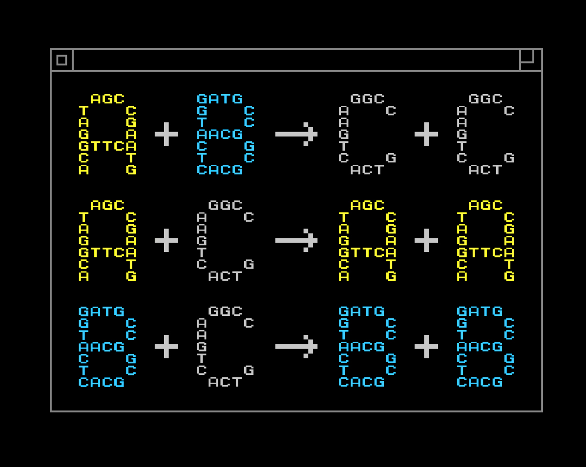
An example of a chemical program. Here, A, B and C are different chemical species. (Credit: Yan Liang/L2XY2.com)
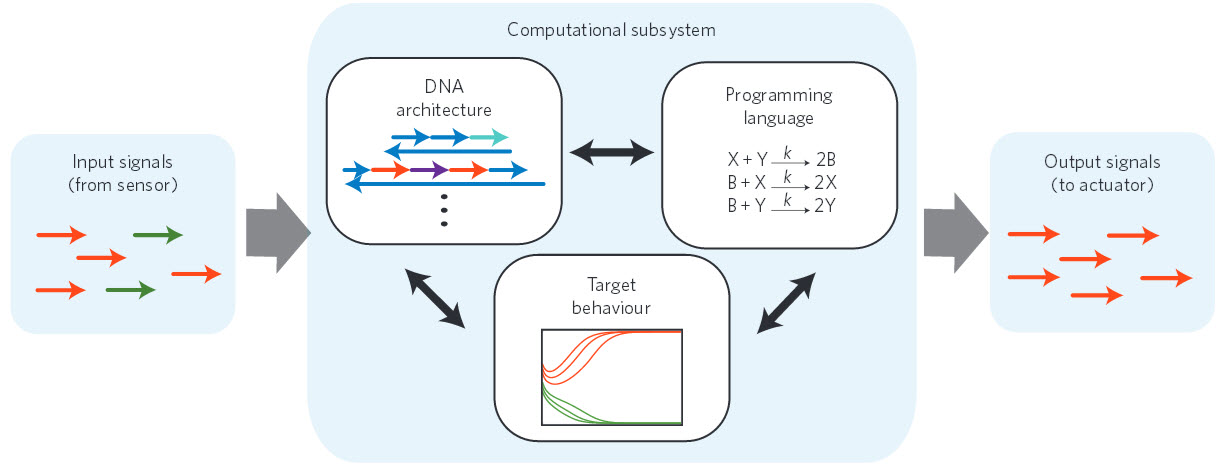
A team led by the University of Washington has developed a programming language to help design chemical-reaction networks (equations that describes how mixtures of chemicals behave).
The objective is to control how DNA molecules build custom-designed molecules in a test tube or cell, which could serve as “smart” drug deliverers or disease detectors at the cellular level, for example.
“We start from an abstract mathematical description of a chemical system, and then use DNA to build the molecules that realize the desired dynamics,” said corresponding author Georg Seelig, a UW assistant professor of electrical engineering and of computer science and engineering.
Currently, when a biologist or chemist makes a certain type of molecular network, the engineering process is complex, cumbersome and hard to repurpose for building other systems.
“I think this is appealing because it allows you to solve more than one problem,” Seelig said. “If you want a computer to do something else, you just reprogram it. This project is very similar in that we can tell chemistry what to do.”
Humans and other organisms already have complex networks of nano-sized molecules that help to regulate cells and keep the body in check. Scientists now are finding ways to design synthetic systems that behave like biological ones with the hope that synthetic molecules could support the body’s natural functions. To that end, a system is needed to create synthetic DNA molecules that vary according to their specific functions.
[+]
Nucleic acid nanocontroller. A standardized signaling protocol based on short single strands of DNA enables the components of the nanocontroller to communicate with each other. The formalism of chemical-reaction networks serves as a programming language that specifies the desired behavior for the computational subsystem. The target behavior is experimentally realized by the DNA architecture. (Credit: Yuan-Jyue Chen et al., Nature Nanotechnology)
The new approach isn’t ready to be applied in the medical field, but future uses could include using this framework to make molecules that self-assemble within cells and serve as “smart” sensors. These could be embedded in a cell, then programmed to detect abnormalities and respond as needed, perhaps by delivering drugs directly to those cells.
Seelig and colleague Eric Klavins, a UW associate professor of electrical engineering, recently received $2 million from the National Science Foundation as part of a national initiative to boost research in molecular programming. The new language will be used to support that larger initiative, Seelig said. The research was also funded by the Burroughs Wellcome Fund and the National Centers for Systems Biology.
California Institute of Technology; Microsoft Research, and University of California, San Francisco researchers were co-authors of the study.

Abstract of Nature Nanotechnology paper:
Biological organisms use complex molecular networks to navigate their environment and regulate their internal state. The development of synthetic systems with similar capabilities could lead to applications such as smart therapeutics or fabrication methods based on self-organization. To achieve this, molecular control circuits need to be engineered to perform integrated sensing, computation and actuation. Here we report a DNA-based technology for implementing the computational core of such controllers. We use the formalism of chemical reaction networks as a ’programming language’ and our DNA architecture can, in principle, implement any behaviour that can be mathematically expressed as such. Unlike logic circuits, our formulation naturally allows complex signal processing of intrinsically analogue biological and chemical inputs. Controller components can be derived from biologically synthesized (plasmid) DNA, which reduces errors associated with chemically synthesized DNA. We implement several building-block reaction types and then combine them into a network that realizes, at the molecular level, an algorithm used in distributed control systems for achieving consensus between multiple agents.

(¯`*• Global Source and/or more resources at http://goo.gl/zvSV7 │ www.Future-Observatory.blogspot.com and on LinkeIn Group's "Becoming Aware of the Futures" at http://goo.gl/8qKBbK │ @SciCzar │ Point of Contact: www.linkedin.com/in/AndresAgostini
Why glial cells should be included in the BRAIN initative

October 2, 2013
[+]
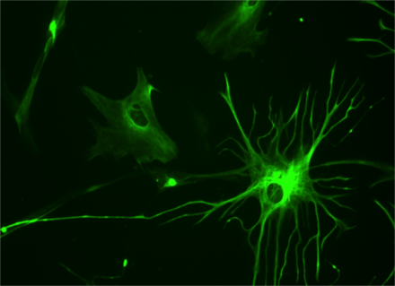
23 week fetal brain culture astrocyte, a type of glial cell (credit: Wikimedia Commons)
Glia, the non-neuronal cells that make up most of the brain, must not be left out of the Brain Research through Advancing Innovative Neurotechnologies (BRAIN) Initiative, says R. Douglas Fields, chief of the Nervous System Development and Plasticity Section at NIH, in Nature News.
“A major stumbling block is the project’s failure to consider that although the human brain contains roughly 100 billion neurons, it contains billions more non-electrical brain cells called glia,” he said.
“These reside outside the neuronal “connectome” and operate beyond the reach of tools designed to probe electrical signaling in neurons. Dismissed as connective tissue when they were first described in the mid-1800s, glia have long been neglected in the quest to understand neuronal signalling. …
“Research is revealing that glia can sense neuronal activity and control it. Various studies also indicate that glia operate in diverse mental processes, for instance, in the formation of memories. They have a central role in brain injury and disease, and they are even at the root of various disorders — such as schizophrenia and Alzheimer’s — previously presumed to be exclusively neuronal. …
“When experts on neuronal plasticity and computational neuroscience came together with glial experts at a workshop in February entitled Glial Biology in Learning and Cognition, held at the US National Science Foundation in Arlington, Virginia, our unanimous conclusion was that neurons working alone provide only a partial explanation for complex cognitive processes, such as the formation of memories.
“The complex branching structure of glial cells and their relatively slow chemical (as opposed to electrical) signalling in fact make them better suited than neurons to certain cognitive processes. These include processes requiring the integration of information from spatially distinct parts of the brain, such as learning or the experiencing of emotions, which take place over hours, days and weeks, not in milliseconds or seconds.”


(¯`*• Global Source and/or more resources at http://goo.gl/zvSV7 │ www.Future-Observatory.blogspot.com and on LinkeIn Group's "Becoming Aware of the Futures" at http://goo.gl/8qKBbK │ @SciCzar │ Point of Contact: www.linkedin.com/in/AndresAgostini
Why don’t we have fusion yet?

October 2, 2013
[+]
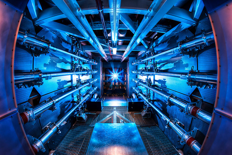
The preamplifiers of the National Ignition Facility. The unified lasers deliver 1.8 megajoules of energy and 500 terawatts of power — 1,000 times more than the United States uses at any one moment. (Credit: Damien Jemison/LLNL)
The dream of igniting a self-sustained fusion reaction with high yields of energy, a feat likened to creating a miniature star on Earth, is getting closer to becoming reality, according the authors of a new review article in the journal Physics of Plasmas.
Researchers at the National Ignition Facility (NIF) report that while there is at least one significant obstacle to overcome before achieving the highly stable precisely directed implosion required for ignition, they have met many of the demanding challenges leading up to that goal since experiments began in 2010.
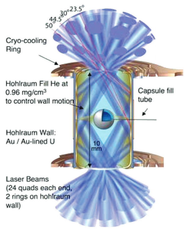
To reach ignition (defined as the point at which the fusion reaction produces more energy than is needed to initiate it), the NIF focuses 192 laser beams simultaneously in billionth-of-a-second pulses inside a cryogenically cooled hohlraum (from the German word for “hollow room”), a hollow cylinder the size of a pencil eraser.
Within the hohlraum is a ball-bearing-size capsule containing two hydrogen isotopes, deuterium and tritium (D-T). The unified lasers deliver 1.8 megajoules of energy and 500 terawatts of power — 1,000 times more than the United States uses at any one moment — to the hohlraum, creating an “X-ray oven” that implodes the D-T capsule to temperatures and pressures similar to those found at the center of the sun.
“What we want to do is use the X-rays to blast away the outer layer of the capsule in a very controlled manner.
That’s so the D-T pellet is compressed to just the right conditions to initiate the fusion reaction,” explained John Edwards, NIF associate director for inertial confinement fusion and high-energy-density science.
One major hurdle
[+]
Schematic of NIF ignition target and capsule (credit: M. J. Edwards et al., Physics of Plasmas)
“In our new review article, we report that the NIF has met many of the requirements believed necessary to achieve ignition — sufficient X-ray intensity in the hohlraum, accurate energy delivery to the target and desired levels of compression — but that at least one major hurdle remains to be overcome: the premature breaking apart of the capsule.”
In the article, Edwards and his colleagues discuss how they are using diagnostic tools developed at NIF to determine likely causes for the problem. “In some ignition tests, we measured the scattering of neutrons released and found different strength signals at different spots around the D-T capsule,” Edwards said.
“This indicates that the shell’s surface is not uniformly smooth and that in some places, it’s thinner and weaker than in others. In other tests, the spectrum of X-rays emitted indicated that the D-T fuel and capsule were mixing too much — the results of hydrodynamic instability — and that can quench the ignition process.”
Edwards said that the team is concentrating its efforts on NIF to define the exact nature of the instability and use the knowledge gained to design an improved, sturdier capsule. Achieving that milestone, he said, should clear the path for further advances toward laboratory ignition.
The project is  led by the Department of Energy’s Lawrence Livermore National Laboratory and includes partners from the University of Rochester’s Laboratory for Laser Energetics, General Atomics, Los Alamos National Laboratory, Sandia National Laboratory, and the Massachusetts Institute of Technology.
The article, “Progress toward ignition on the National Ignition Facility” by M.J. Edwards et al. appears in the journal Physics of Plasmas. Authors of this paper are affiliated with Lawrence Livermore National Laboratory, General Atomics in San Diego, Calif., the Massachusetts Institute of Technology, the University of Rochester, Los Alamos National Laboratory, and Sandia National Laboratory.
Physics of Plasmas, produced by AIP Publishing with the cooperation of the American Physical Society (APS) Division of Plasma Physics, is devoted to original experimental and theoretical contributions to the physics of plasmas.


(¯`*• Global Source and/or more resources at http://goo.gl/zvSV7 │ www.Future-Observatory.blogspot.com and on LinkeIn Group's "Becoming Aware of the Futures" at http://goo.gl/8qKBbK │ @SciCzar │ Point of Contact: www.linkedin.com/in/AndresAgostini
Extending MRI to nanoscale resolution

October 2, 2013
[+]
Illustration of the experimental setup shows the two unique components of the team’s novel MRI technique that was successful in producing a 2D MRI image with spatial resolution on the nanoscale (credit: Budakian et al./University of Illinois at Urbana-Champaign)
University of Illinois at Urbana-Champaign and Northwestern University researchers have devised a novel nuclear magnetic resonance imaging (MRI) technique that delivers about 10­ nanometers spatial resolution.
This represents a significant advance in MRI sensitivity. Current MRI techniques commonly used in medical imaging yield spatial resolutions on the millimeter length scale, with the highest-resolution experimental instruments giving spatial resolution of a few micrometers.
“Our approach brings MRI one step closer in its eventual progress toward atomic-scale imaging,” said U. of I. physicist Raffi Budakian, who led the research effort.
The new breakthrough technique introduces two unique components to overcome obstacles to applying classic pulsed magnetic resonance techniques in nanoscale systems:
  • A novel protocol for spin manipulation applies periodic radio-frequency magnetic field pulses to encode temporal correlations in the statistical polarization of nuclear spins in the sample.
  • A nanoscale metal constriction focuses current, generating intense magnetic-field pulses.
In their proof-of-principle demonstration, the team used an ultrasensitive magnetic resonance sensor based on a silicon nanowire oscillator to reconstruct a two-dimensional projection image of the proton density in a polystyrene sample at nanoscale spatial resolution.
“We expect this new technique to become a paradigm for nanoscale magnetic-resonance imaging and spectroscopy into the future,” added Budakian. “It is compatible with and can be incorporated into existing conventional MRI technologies.”
Exclusive KurzweilAI interview with Prof. Budakian
What are the major applications of your research and when can we expect to see operational devices?
Specifically, we are focused on imaging biological systems between 1–100 nm. These include proteins and viruses. MRI is a powerful tool for studying biological systems because it offers a host of unique modalities for imaging. It is nondestructive, fully three-dimensional, and chemically specific. Extending these capabilities to the nanometer scale would, among other things, transform our understanding of protein structure, which would enable more effective drug development. Our approach would permit the application of established techniques in clinical MRI to the nanometer scale.
We are in the beginning stages of this new technology. We need several more years of technique development before we can apply this technique to answer biologically relevant questions. Of course, the speed of progress depends a great deal on the funding situation. The application of this technique will not be in a clinical setting. I have not approached any commercial companies with this idea. It’s still very new.
What is the highest resolution available in current MRI devices and how do they compare with your work?
The highest resolution inductively-coupled MRI measurements that I am aware of is 3.7 x 3.3 x 3.3 micrometers [2]. There  is a number of people in the force-detected MRI community working on developing nanoscale MRI.. There is also a growing community of people trying to apply nitrogen vacancy centers to nanoscale MRI [3,4].
The first work that demonstrated nanoscale MRI imaging was by Degan et al.[5]. Like the previous work, our approach uses force-detected magnetic resonance imaging. Our approach differs in several important aspects to that work. One of the most significant differences is the use of time-dependent magnetic field gradients for spin detection and imaging. The ability to control the time dependence of the magnetic fields permits the use of all other pulsed magnetic resonance techniques for nanoscale imaging and spectroscopy.
What are your plans for future development?
The goal of our work is to extend the capabilities of MRI to the nanometer scale. In this initial proof-of-concept work, we demonstrated 10-nm spatial resolution imaging of proton spins in polystyrene. In the next 2–3 years, our goal is to demonstrate proton spin imaging in biological systems with 1–3 nm spatial resolution.

Abstract of Physical Review X paper [1]

We report a method for nanometer-scale pulsed nuclear magnetic resonance imaging and spectroscopy. Periodic radio-frequency pulses are used to create temporal correlations in the statistical polarization of a solid organic sample. The spin density is spatially encoded by applying a series of intense magnetic field gradient pulses generated by focusing electric current through a nanometer-scale metal constriction. We demonstrate this technique using a silicon nanowire mechanical oscillator as a magnetic resonance sensor to image 1H spins in a polystyrene sample. We obtain a two-dimensional projection of the sample proton density with approximately 10-nm resolution.

(¯`*• Global Source and/or more resources at http://goo.gl/zvSV7 │ www.Future-Observatory.blogspot.com and on LinkeIn Group's "Becoming Aware of the Futures" at http://goo.gl/8qKBbK │ @SciCzar │ Point of Contact: www.linkedin.com/in/AndresAgostini
Tuesday, October 1, 2013


How to make ceramics that bend without breaking

New materials developed at MIT could lead to actuators on a chip and self-deploying medical devices
October 1, 2013
[+]
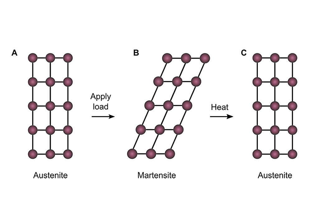
When subjected to a load, the molecular structure of a zirconia ceramic material (austenite) deforms (in its martensite phase) rather than cracking,. When heated, it then returns to its original shape. (Credit: Lai et al./MIT)
Ceramics tend to crack under stress. But researchers from MIT and Nanyang Technological University in Singapore have developed a way of making minuscule flexible ceramic objects that also have a “memory” for shape (when bent and then heated, they return to their original shapes).
The surprising discovery is reported this week in the journal Science, in a paper by MIT graduate student Alan Lai, professor Christopher Schuh, and two collaborators in Singapore.
Shape-memory materials, which can bend and then snap back to their original configurations in response to a temperature change, have been known since the 1950s in metals, and some polymers, but not in ceramics,” explains Schuh, the Danae and Vasilis Salapatas Professor of Metallurgy and head of MIT’s Department of Materials Science and Engineering.
The team accomplished this in two key ways.
  • Created tiny ceramic objects, invisible to the naked eye: “When you make things small, they are more resistant to cracking,” Schuh says.
  • Made the individual crystal grains span the entire small-scale structure, removing the crystal-grain boundaries where cracks are most likely to occur.
Those tactics resulted in tiny samples of ceramic material with deformability equivalent to about 7 percent of their size. “Usually if you bend a ceramic by 1 percent, it will shatter,” Schuh says. But these tiny filaments, with a diameter of just 1 micrometer — one millionth of a meter — can be bent by 7 to 8 percent repeatedly without any cracking, he says.
Ceramic-like strength, but metal-like ductility
These materials could be important tools for those developing micro- and nanodevices, such as for biomedical applications, Schuh says, such as microactuators to trigger actions within such devices — the release of drugs from tiny implants, for example.
Compared to the materials currently used in microactuators, Schuh says, the strength of the ceramic would allow it to exert a stronger push in a microdevice.
The ceramics used in this research were made of zirconia, but the same techniques should apply to other ceramic materials. Zirconia is “one of the most well-studied ceramics,” Lai says, and is already widely used in engineering. It is also used in fuel cells, considered a promising means of providing power for cars, homes and even for the electric grid. While there would be no need for elasticity in such applications, the material’s flexibility could make it more resistant to damage.
The material combines some of the best attributes of metals and ceramics, the researchers say: Metals have lower strength but are very deformable, while ceramics have much greater strength, but almost no ductility — the ability to bend or stretch without breaking. The newly developed ceramics, Schuh says, have “ceramic-like strength, but metal-like ductility.”

(¯`*• Global Source and/or more resources at http://goo.gl/zvSV7 │ www.Future-Observatory.blogspot.com and on LinkeIn Group's "Becoming Aware of the Futures" at http://goo.gl/8qKBbK │ @SciCzar │ Point of Contact: www.linkedin.com/in/AndresAgostini
NASA space telescopes find patchy clouds on exotic world

October 1, 2013
kepler-7b
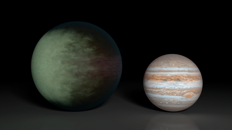
Kepler-7b (left), which is 1.5 times the radius of Jupiter (right), is the first exoplanet to have its clouds mapped. This artist’s concept shows what those clouds might look like. The cloud map was produced using data from NASA’s Kepler and Spitzer space telescopes. The map shows that clouds cover the western side of the gaseous planet, leaving the east cloud-free. Researchers speculate the clouds are made up of minerals containing silicates. Kepler-7b is one of the puffiest, or least dense, planets known. While it is 1.5 times the size of Jupiter is has only about half the mass. (Credit: NASA/JPL-Caltech/MIT)
Astronomers using data from NASA’s Kepler and Spitzer space telescopes have created the first cloud map of a planet beyond our solar system: a sizzling, Jupiter-like world known as Kepler-7b.
The planet is marked by high clouds in the west and clear skies in the east. Previous studies from Spitzer have resulted in temperature maps of planets orbiting other stars, but this is the first look at cloud structures on a distant world.
“By observing this planet with Spitzer and Kepler for more than three years, we were able to produce a very low-resolution ‘map’ of this giant, gaseous planet,” said Brice-Olivier Demory of MIT. Demory is lead author of a paper accepted for publication in the Astrophysical Journal Letters. “We wouldn’t expect to see oceans or continents on this type of world, but we detected a clear, reflective signature that we interpreted as clouds.”
Kepler has discovered more than 150 exoplanets, which are planets outside our solar system, and Kepler-7b was one of the first. The telescope’s problematic reaction wheels prevent it from hunting planets any more, but astronomers continue to pore over almost four years’ worth of collected data.
Kepler’s visible-light observations of Kepler-7b’s moon-like phases led to a rough map of the planet that showed a bright spot on its western hemisphere. But these data were not enough on their own to decipher whether the bright spot was coming from clouds or heat. The Spitzer Space Telescope played a crucial role in answering this question.
Like Kepler, Spitzer can fix its gaze at a star system as a planet orbits around the star, gathering clues about the planet’s atmosphere. Spitzer’s ability to detect infrared light means it was able to measure Kepler-7b’s temperature, estimating it to be between 1,500 and 1,800 degrees Fahrenheit (1,100 and 1,300 Kelvin).
This is relatively cool for a planet that orbits so close to its star — within 0.06 astronomical units (one astronomical unit is the distance from Earth and the sun) — and, according to astronomers, too cool to be the source of light Kepler observed. Instead, they determined, light from the planet’s star is bouncing off cloud tops located on the west side of the planet.
“Kepler-7b reflects much more light than most giant planets we’ve found, which we attribute to clouds in the upper atmosphere,” said Thomas Barclay, Kepler scientist at NASA’s Ames Research Center in Moffett Field, Calif. “Unlike those on Earth, the cloud patterns on this planet do not seem to change much over time — it has a remarkably stable climate.”
A new tool for studying exoplanets similar to Earth
The findings are an early step toward using similar techniques to study the atmospheres of planets more like Earth in composition and size.
“With Spitzer and Kepler together, we have a multi-wavelength tool for getting a good look at planets that are trillions of miles away,” said Paul Hertz, director of NASA’s Astrophysics Division in Washington. “We’re at a point now in exoplanet science where we are moving beyond just detecting exoplanets, and into the exciting science of understanding them.”
Kepler identified planets by watching for dips in starlight that occur as the planets transit, or pass in front of their stars, blocking the light. This technique and other observations of Kepler-7b previously revealed that it is one of the puffiest planets known: if it could somehow be placed in a tub of water, it would float. The planet was also found to whip around its star in just less than five days.
Explore all 900-plus exoplanet discoveries with NASA’s “Eyes on Exoplanets,” a fully rendered 3D visualization tool, available for download. The program is updated daily with the latest findings from NASA’s Kepler mission and ground-based observatories around the world as they search for planets like our own.
University of California, Santa Cruz, California Institute of Technology, the University of Bern, Yale University, the University of Liège, the French National Center for Scientific Research, and Northwestern University were also involved in the research.


(¯`*• Global Source and/or more resources at http://goo.gl/zvSV7 │ www.Future-Observatory.blogspot.com and on LinkeIn Group's "Becoming Aware of the Futures" at http://goo.gl/8qKBbK │ @SciCzar │ Point of Contact: www.linkedin.com/in/AndresAgostini
NASA wants investigations for a Mars 2020 rover

October 1, 2013
mars_2020
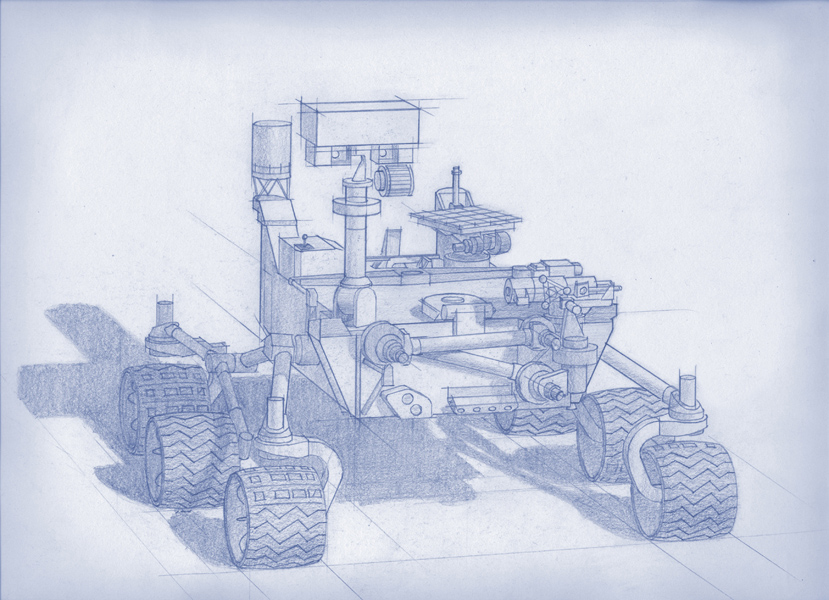
Planning for NASA’s 2020 Mars rover envisions a basic structure that capitalizes on the design and engineering work done for the NASA rover Curiosity, which landed on Mars in 2012, but with new science instruments selected through competition for accomplishing different science objectives (credit: NASA/JPL-Caltech)
NASA has announced an open competition for the planetary community to submit proposals for the science and exploration technology instruments that would be carried aboard the agency’s next Mars rover, scheduled for launch in July/August of 2020.
The Mars 2020 rover will explore and assess Mars as a potential habitat for life, search for signs of past life, collect carefully selected samples for possible future return to Earth, and demonstrate technology for future human exploration of the Red Planet.
Officially called the Mars 2020 Mission Investigations Announcement of Opportunity (AO), this competition solicits flight investigations for which each principal investigator or scientist is responsible for a complete space flight investigation, including instrument hardware, mission operations and data analysis. T
he total allocated cost for development of all the investigations selected and funded by NASA is approximately $130 million.
The competitively selected instruments will be placed on a rover similar to Curiosity, which landed on Mars in August 2012. Using Curiosity’s design will help minimize mission costs and risks and deliver a rover that can accomplish the mission objectives. The Mars 2020 mission also would build upon the scientific accomplishments of Curiosity and other previous Mars missions.
What’s different about Mars 2020
In January 2013, NASA appointed a Science Definition Team to outline objectives for the Mars 2020 mission. The team, composed of 19 scientists and engineers from universities and research organizations, proposed a mission concept that could accomplish several high-priority planetary science goals and be a major step in meeting President Obama’s challenge to send humans to Mars in the 2030s.
According to the Science Definition Team, looking for signs of past life is the next logical step.
“The Mars 2020 mission will provide a unique capability to address the major questions of habitability and life in the solar system,” said Jim Green, director of NASA’s Planetary Science Division in Washington. “The science conducted by the rover’s instruments also would expand our knowledge of Mars and provide the context needed to make wise decisions about whether to return any collected samples to Earth.”
This rover will make measurements of mineralogy and rock chemistry down to a microscopic scale, so that we might be able to understand the Martian environment surrounding the rover’s landing site and identify evidence of possible past life.
The 2020 rover could also make measurements and conduct technology demonstrations to help designers of a human expedition understand any hazards posed by Martian dust and demonstrate how to collect carbon dioxide, which could be a resource for making oxygen and rocket fuel.
“The Mars 2020 rover will test technologies that are key to one-day landing human explorers on the Red Planet,” said Jason Crusan, director of NASA’s Advanced Exploration Systems Division. “New technologies could allow astronauts to live off the land as they explore the ancient valleys of Mars. The capability to manufacture breathable air, rocket fuel, water and more may forever change how we explore space.”


(¯`*• Global Source and/or more resources at http://goo.gl/zvSV7 │ www.Future-Observatory.blogspot.com and on LinkeIn Group's "Becoming Aware of the Futures" at http://goo.gl/8qKBbK │ @SciCzar │ Point of Contact: www.linkedin.com/in/AndresAgostini
A hidden genetic code for better designer genes

How rare "words" in bacterial genes boost protein production
October 1, 2013
Codon
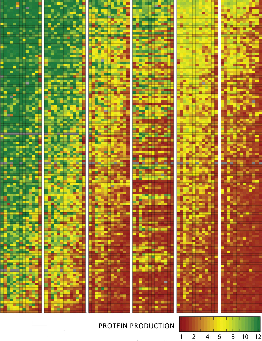
Wyss Institute scientists synthesized 14,000 snippets of DNA (individual pixels) and tested how well they promote protein synthesis. They found that those with rare ³words² (codons) near the start of bacterial genes made more protein (green pixels) by removing roadblocks to protein production. (Credit: Wyss Institute)
Scientists routinely seek to reprogram bacteria to produce proteins for drugs, biofuels and more, but they have struggled to get those bacteria to follow orders.
A hidden feature of the genetic code, it turns out, could achieve that.  The feature controls how much of the desired protein bacteria produce, a team from the Wyss Institute for Biologically Inspired Engineering at Harvard University reported in the September 26 online issue of Science.
Rare condons
The researchers combined high-speed “next-generation” DNA sequencing and DNA synthesis technologies and found that using more rare words, or codons, near the start of a gene removes roadblocks to protein production.
The research team is headed by Sri Kosuri, Ph.D., a Wyss Institute staff scientist, George Church, Ph.D., a core faculty member at the Wyss Institute and professor of genetics at Harvard Medical School, and Daniel Goodman, a Wyss Institute graduate research fellow,
“Now that we understand how rare codons control gene expression, we can better predict how to synthesize genes that make enzymes, drugs, or whatever you want to make in a cell,” Kosuri said.
The findings could be a boon for biotechnologists,and could help synthetic biologists reprogram bacteria to make new drugs and biological devices.
To produce a protein, a cell must first make working copies of the gene encoding it. These copies, called messenger RNA (mRNA), consist of a specific string of words, or codons. Each codon represents one of the 20 different amino acids that cells use to assemble proteins. But since the cell uses 61 codons to represent 20 amino acids, many codons have synonyms that represent the same amino acid.
In bacteria, as in books, some words are used more often than others, and molecular biologists have noticed over the last few years that rare codons appear more frequently near the start of a gene. What’s more, genes whose opening sequences have more rare codons produce more protein than genes whose opening sequences do not.
No one knew for sure why rare codons had these effects, but many biologists suspected that they function as a highway on-ramp for ribosomes, the molecular machines that build proteins. According to this idea, called the codon ramp hypothesis, ribosomes wait on the on-ramp, then accelerate slowly along the mRNA highway, allowing the cell to make proteins with all deliberate speed.
But without the on-ramp, the ribosomes gun it down the mRNA highway, then collide like bumper cars, causing traffic accidents that slow protein production. Other biologists suspected rare codons acted via different mechanisms. These include mRNA folding, which could create roadblocks for ribosomes that block the highway and slow protein production.
A high-speed, multiplexed method 
To see which ideas were correct, the three researchers used a high-speed, multiplexed method that they’d reported in August in The Proceedings of the National Academy of Sciences.
First, they tested how well rare codons activate genes by mass-producing 14,000 snippets of DNA with either common or rare codons; splicing them near the start of a gene that makes cells glow green, and inserting each of those hybrid genes into different bacteria. Then they grew those bugs, sorted them into bins based on how intensely they glowed, and sequenced the snippets to look for rare codons.
They found that genes that opened with rare codons consistently made more protein, and a single codon change could spur cells to make 60 times more protein.
“That’s a big deal for the cell, especially if you want to pump out a lot of the protein you’re making,” Goodman said.
The results were also consistent with the codon-ramp hypothesis, which predicts that rare codons themselves, rather than folded mRNA, slow protein production. But the researchers also found that the more mRNA folded, the less of the corresponding protein it produced — a result that undermined the hypothesis.
To put the hypothesis to a definitive test, the Wyss team made and tested more than 14,000 mRNAs — including some with rare codons that didn’t fold well, and others that folded well but had no rare codons. By quickly measuring protein production from each mRNA and analyzing the results statistically, they could separate the two effects.
The results showed clearly that RNA folding, not rare codons, controlled protein production, and that scientists can increase protein production by altering folding, Goodman said.
The new method could help resolve other thorny debates in molecular biology. “The combination of high-throughput synthesis and next-gen sequencing allows us to answer big, complicated questions that were previously impossible to tease apart,” Church said.
“These findings on codon use could help scientists engineer bacteria more precisely than ever before, which is tremendous in itself, and they provide a way to greatly increase the efficiency of microbial manufacturing, which could have huge commercial value as well,” said Wyss Institute Founding Director Don Ingber, M.D., Ph.D. “They also underscore the incredible value of the new automated technologies that have emerged from the Synthetic Biology Platform that George leads, which enable us to synthesize and analyze genes more rapidly than ever before.”
The work was funded by the Department of Energy, the Office of Naval Research, Agilent Technologies, an NSF Graduate Research Fellowship to Daniel Goodman, and the Wyss Institute.


(¯`*• Global Source and/or more resources at http://goo.gl/zvSV7 │ www.Future-Observatory.blogspot.com and on LinkeIn Group's "Becoming Aware of the Futures" at http://goo.gl/8qKBbK │ @SciCzar │ Point of Contact: www.linkedin.com/in/AndresAgostini

Human on a chip

October 1, 2013
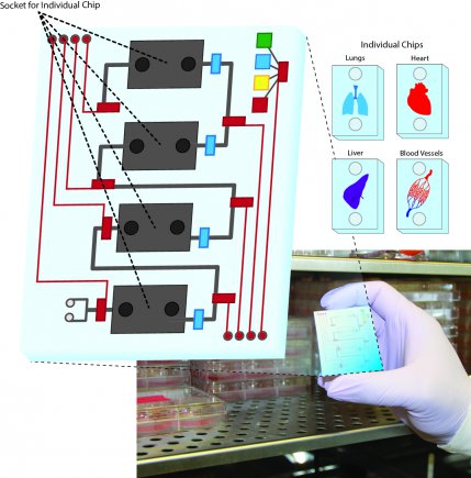
(Credit: U.S. Army)
Army scientists at the Edgewood Chemical Biological Center (ECBC) and academic collaborators are conducting research on organoids (small swatches of human tissue) on microchips.
The “human on a chip” research focuses on in vitro human organ constructs (for the heart, liver, lung and the circulatory system) in communication with each other. The goal is to assess effectiveness and toxicity of drugs in a way that is relevant to humans and their ability to process these drugs.
“The screening models will be used to assess the efficacy and safety of medical mitigation procedures and countermeasures for the soldier and the nation as a whole,” said Dr. Harry Salem, ECBC’s chief scientist for Life Sciences.
Each organ-on-a-chip is about the size of a thumb drive and is an “organoid” (a structure that resembles an organ in appearance or function), designed to mimic the properties of an actual human organ.
The organoids are created by induced pluripotent stem cells made from adult skin cells. They comprise multiple layers of cells growing on a membrane, connected to each other by microfluidics (tiny micro channels) that copy the function of blood vessels.
Their primary purpose is to take the place of animal research. According to Salem, compounds quite often behave differently in people than they do in animals. For that reason, human-estimate studies are used, but do not always accurately reflect the human response. Due to the species-specific differences by which compounds are metabolized, a drug tested on a laboratory rat doesn’t always translate well to a human.
In some cases, no animal testing can mimic the human response. Asthma, for example, is a uniquely human disease. Since human-on-a-chip is made from human cells, it is the next best thing. Human tissue reacts like human tissue.
New predictive models of toxicity
The researchers anticipate that new predictive models of toxicity will result from the more accurate human-on-a-chip testing, saving time and money. Pharmaceuticals tested on animals fail to work on humans 90 percent of the time. This technology will result in fewer test failures. Scientists will be able to narrow their research efforts by identifying which therapeutics will be effective or fail early on in the testing process, the researchers say.
The center houses the only laboratories in the United States that the Chemical Weapons Convention permits to produce chemical warfare agent for testing purposes. ECBC will test the human-on-a-chip against chemical warfare agents to learn more about how the body will respond to agent exposure and explore various treatment options for exposures.
“The human-on-a-chip promises to accelerate the pace of research and consequently scientific breakthroughs,” Dr. Russell Dorsey, a research microbiologist and one of the members performing the in vitro testing at ECBC, said. “For the military, our human-on-a-chip research will save actual warfighters’ lives.”
The center will be collaborating with the U.S. Army Medical Research Institute of Chemical Defense, Wake Forest, Harvard, and the University of Michigan on the chip design.
The five-year research project is funded by the Defense Threat Reduction Agency.

(¯`*• Global Source and/or more resources at http://goo.gl/zvSV7 │ www.Future-Observatory.blogspot.com and on LinkeIn Group's "Becoming Aware of the Futures" at http://goo.gl/8qKBbK │ @SciCzar │ Point of Contact: www.linkedin.com/in/AndresAgostini
Monday, September 30, 2013


Syria's War Viewed Almost in Real Time

For the first time, the war in Syria has given global audiences a close-up view of war

    By
  • MELIK KAYLAN
The proliferation of cellphone cams and social media have produced, via the instant upload, a new phenomenon: the first YouTube war. Melik Kaylan explains the impact of online videos in the Syrian conflict.
In one video, two men in jeans and hoodies take a rocket tube to a rooftop and fire it. From another angle, we see three Syrian tanks in a row. The middle one takes a huge hit, emits a sheet of orange flame and burns away; a smoldering figure jumps from the tank and runs off as bullets smack the ground around him.
Another video opens with a long take of a busy traffic area, full of buses and pedestrians, some of whom seem to be soldiers. One bus suddenly erupts from within, and dozens of people collapse. The videotaker repeatedly cries "Allahu akbar" and claims to be from the Al Nusra Front, a radical Islamist group.
Syria Baynetna/YouTube
A screengrab from a YouTube video from September
The unprecedented confluence of two technologies—cellphone cams and social media—has produced, via the instant upload, a new phenomenon: the YouTube war. For the first time in history, the extended war in Syria has furnished global audiences with a sofa-side view of what it feels like to be there, almost in real time.
Since January 2012, according to official YouTube figures, over a million videos have been uploaded, with hundreds of millions of views to date. The company doesn't, as a matter of policy, delete even very graphic videos that are news-oriented, but it does sometimes append warnings. Bus bombs, firefights, raw scenes of bloodshed and tragedy, interrogations and executions, tank kills, deaths by sniper—the full brutal spectrum of real-life combat drama is thus now on display at the click of a finger.
islamtv001/YouTube
A screengrab from a YouTube video from July
Videos have poured in from all sides of the war in Syria, part of a social-media struggle to document events and ultimately to influence them. According to Hassan, a young Syrian documentary filmmaker who moved last month to the U.S. to join family and to avoid the draft (his name is changed here to prevent identification), "virtually every neighborhood, for or against Assad, has a media center that documents and processes what happens. Many went abroad for training, with funds from outside or state subsidy."
On YouTube, even those who don't know Arabic can detect the biases. Content against Syrian President Bashar al-Assad often gets captioned in English and trumpets the successes of the rebel Free Syrian Army. Wins for Mr. Assad's forces are categorized under the heading Syrian Arab Army. Troops in uniform, including the Iran-backed militia Hezbollah, tend to be pro-Assad. Rebel groups like the al-Qaeda-linked Al Nusra Front and the Islamic State of Iraq and Al Sham have logos with stylized Islamic scripts (apparently they can afford to outsource for accomplished graphic design). According to Hassan and others, each fighting group now brings along its own more-or-less professional cameramen.
SNN Shaam English News/YouTube
A screengrab from a YouTube video from February
But despite all the expertise in propaganda, the products can often seem bafflingly alienating or repellent. A pro-Assad channel entitled SyriaTube likes to put out close-up scenes of rebels taking hits, collapsing and dying, presented with insouciant music from spaghetti westerns and "Bye Bye" in large letters. The producers don't seem to sense anything discordant or sadistic in the juxtaposition.
One wonders also how they get hold of videos that had to be originally shot by rebels showing one of their own being hit. Hassan explains that they find them on prisoners or dead rebels: "The first thing both sides search for is cellphones or cameras."
On the rebel side, the incessant cries of Allahu akbar come across, finally, as barbaric because they are uttered so indiscriminately: at the death of a comrade, the destruction of a tank, the execution of a prisoner, the killing of innocents or the launch of a rocket toward the enemy. The implicit suggestion is that God presides as much over their boastful cruelty as over their prayers for mercy for the souls of the dead.
An Al Jazeera editor who has worked in the region for some years (and wishes to remain anonymous) says that the early videos from the conflict didn't feature such vehement religiosity. "At a certain point, fighters began to produce footage specifically to appeal to Gulf and Saudi sources of funding," he said. It has now reached the point that groups "perform missions for the camera and go back to funders saying, 'This is what $50,000 got you. For $500,000, we could knock out an entire base,' or some such."
This confluence between mercenary motives and killing for the faith isn't news to Ali Soufan, the Lebanese-American former FBI agent who, after the attack on the USS Cole and 9/11, successfully interrogated numerous Al Qaeda operatives, including Osama bin Laden's bodyguard. "Jihadi elements learned years ago to use video and social media for recruitment and funding," he said. "They're in the business of promoting themselves while the mainstream media isn't promoting them. They know it's a kind of theater."
For Mr. Soufan and others who monitor online traffic for antiterror purposes, Syrian war videos have proved a vital resource. Charles Lister, a prominent analyst for the U.K.-based IHS Jane's Terrorism and Insurgency Center, has acknowledged his dependence on videos. In a report over the summer, he analyzed scores of videos to determine the quality and quantity of weapons reaching the rebels. This occurred at a moment of intense public debate about the purported shortfall of Western support for the insurgency.
Truthloader/YouTube
A screengrab from a YouTube video.
The use and misuse of war videos—their secondary and tertiary life out in the ether—will surely become part of our future experience of wars. What clues does the Syria experience offer? According to Hazami Barmada, an Arab-American who works as a consultant to various states in the Middle East, "How the videos are shared, the social media commentary around them, is as important as the content. People in the region are fully engaged in that dimension. There are two conflicts, the war and the digital war, which globalizes it."
What has this wider awareness added up to? "I don't see any good effect," says Marc Ginsberg, the former U.S. ambassador to Morocco. "Where are all the protests against Assad in Arab countries? Instead, it's probably sucked more people into the war from outside."
Videos showing the ghastly toll of the chemical weapons attack in late August proved to be a turning point of sorts, prompting a more focused debate about Western intervention. Up to then, though, Syrians had recorded any number of atrocities for global eyes, with no result. One might conclude that the age of YouTube war will bring the worst of possible outcomes: an ever-growing number of us witnessing horrors while at the same time growing fatalistic about them—just as war victims themselves do.

(¯`*• Global Source and/or more resources at http://goo.gl/zvSV7 │ www.Future-Observatory.blogspot.com and on LinkeIn Group's "Becoming Aware of the Futures" at http://goo.gl/8qKBbK │ @SciCzar │ Point of Contact: www.linkedin.com/in/AndresAgostini